 Bringing Gold-Standard Imaging to the OT
Digital Subtraction Angiography remains the gold standard for diagnosing cerebrovascular conditions such as aneurysms, arteriovenous malformations (AVMs), and vasculitis. Traditionally performed in dedicated catheterization laboratories by interventional radiologists, DSA is critical when CT angiography results are inconclusive or when surgical planning requires real-time vascular detail.
By integrating a mobile 3D C-arm system into the neurosurgery operating theatre, the NIMS team aimed to give neurosurgeons direct control over angiographic imaging. The approach reduces reliance on external cath lab scheduling and enhances intraoperative decision-making.
Study Design and Institutional Framework
The observational study was conducted between October 2023 and January 2025 at NIMS, a tertiary-care government teaching hospital. Institutional approval was obtained before initiating the program.
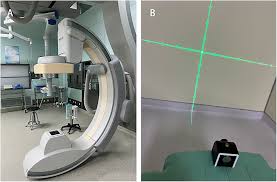
All 100 cases were performed using a multi-axis 3D C-arm system equipped with flat-panel detection and 3D reconstruction capabilities. The operating theatre was modified with radiation shielding, lead-lined doors, and a radiolucent operating table to allow smooth C-arm movement and optimal imaging angles.
Patient Profile and Indications
Among the 100 procedures performed, 98 were diagnostic angiographies and two involved limited endovascular intervention for vasospasm management using intra-arterial nimodipine.
The majority of patients—88%—presented with spontaneous subarachnoid hemorrhage (SAH), requiring detailed vascular evaluation to identify aneurysms or other causes. Additional indications included follow-up imaging in moyamoya disease, evaluation of AVMs, and selected unexplained intracerebral hemorrhages.
The cohort included 48 men and 52 women, with a mean age of 55 years. Aneurysms were identified in 89% of SAH cases.
Technique and Safety Measures
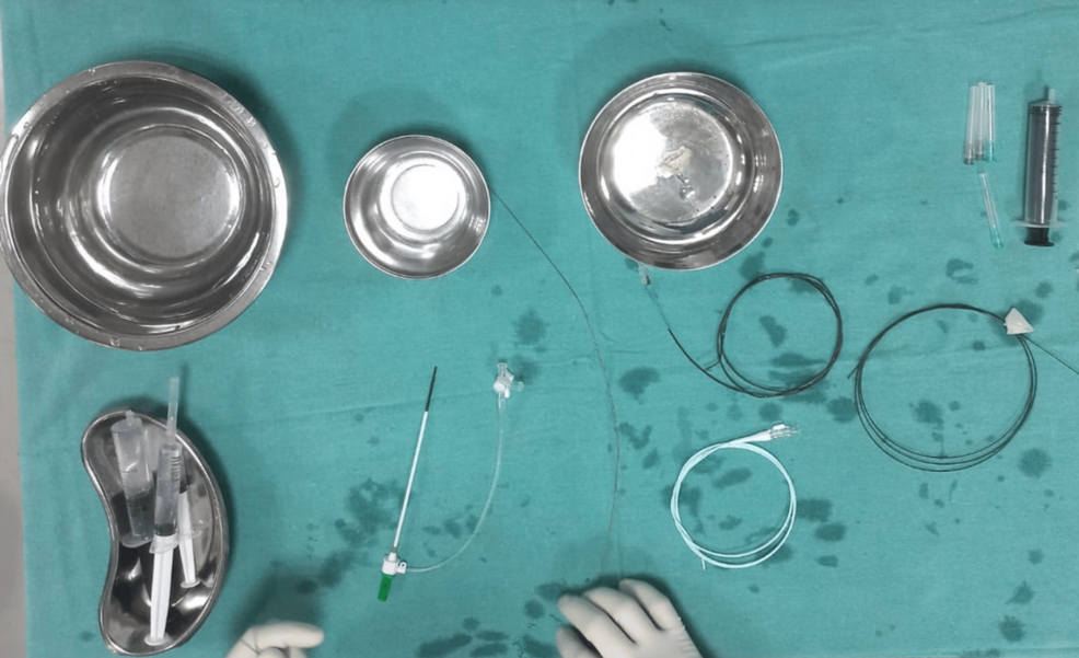
All procedures were performed via right femoral artery access using the Seldinger technique. Selective cannulation of bilateral carotid and vertebral arteries ensured complete evaluation of anterior and posterior circulations.
Standard angiographic projections including anteroposterior, lateral, oblique, and Towne’s views were obtained depending on suspected pathology. Most cases were conducted under local anesthesia, enabling continuous neurological monitoring.
Radiation exposure was minimized following the “As Low As Reasonably Achievable” (ALARA) principle. Fluoroscopy time was carefully controlled, and protective shielding was mandatory for all staff.
Challenges and Outcomes
Five patients exhibited markedly tortuous carotid anatomy, making catheter navigation technically challenging. In two such cases, the procedure was safely abandoned to avoid complications.
Importantly, no major complications such as stroke, access-site hematoma, or mortality were reported across the 100 procedures. Post-procedural care included ICU monitoring, neurological assessments, and access-site surveillance.
The absence of serious adverse events underscores the feasibility and safety of performing DSA within a neurosurgical OT setting when supported by proper training and infrastructure.
A Step Toward Hybrid Neurosurgery
Experts involved in the initiative emphasize that modern neurosurgery is evolving toward a hybrid model, combining open microsurgical and endovascular skills. Neurosurgeons trained in angiography are better equipped to interpret vascular anatomy and manage complications promptly.
The integration of 3D C-arm-based DSA expands real-time intraoperative imaging capabilities without the need for a dedicated hybrid suite. This model may be particularly beneficial in resource-constrained public hospitals.
The NIMS experience demonstrates that with structured planning, training, and safety protocols, neurosurgical DSA programs can be successfully implemented within conventional operating rooms potentially reshaping neurovascular care in India.